Foot Muscles Mri - MRI with user outlined plantar intrinsic and extrinsic ... / Muscles that move the foot and toes.. Abdm, abductor digiti minimi muscle; Please come back soon to see the finished work! 10 foot and ankle craig r. Head, neck, arm, foot, pelvis, etc. Muscles that move the foot and toes.
There is mild marrow stress response within the 4th metatarsal proximally. Muscle damage may cause muscle pain and muscle weakness may cause difficulty lifting the arms above the shoulders, climbing stairs, or arising from a sitting position. Learn about foot and ankle mri here. Head, neck, arm, foot, pelvis, etc. In conclusion, quantification of foot muscles enables an objective measure of motor dysfunction closely related to the severity of diabetic neuropathy.
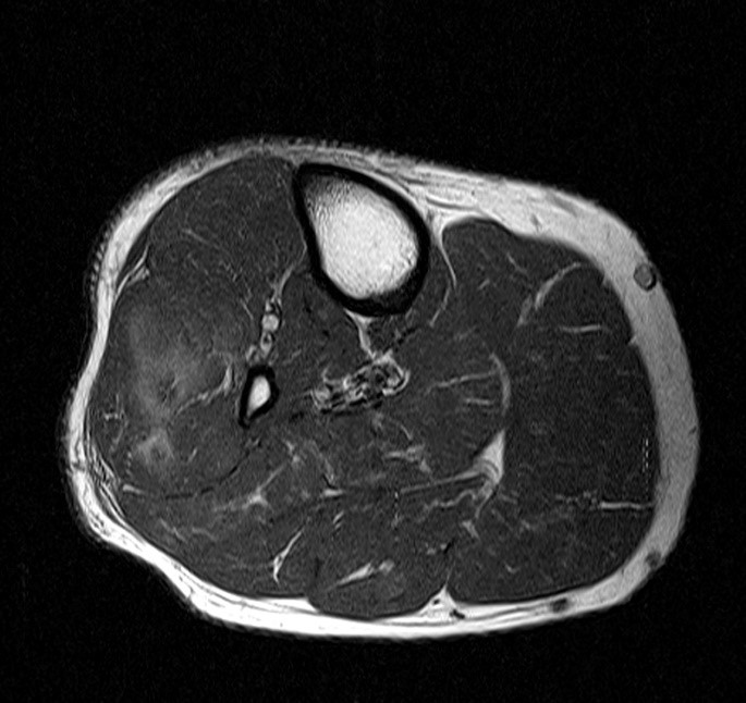
Compartment Syndrome of the Leg - Radsource from radsource.us The deformity of the foot with abnormal pressure distribution on the plantar surface coupled with reduced or loss of sensation, makes the foot. Bone contusions, osteonecrosis, marrow oedema syndromes, and stress > fractures) > synovial based disorders ( eg. They are considered voluntary muscles. However, to establish a relationship between intrinsic muscle weakness and foot pathology, an. Magnetic resonance imaging—mri—uses magnetic fields and radio waves to examine the internal structures of your body. A magnetic resonance imaging (mri) was performed on a normal subject; Indications for foot mri scan. Abdm, abductor digiti minimi muscle;
Abdm, abductor digiti minimi muscle;
Learn about foot and ankle mri here. Muscle damage may cause muscle pain and muscle weakness may cause difficulty lifting the arms above the shoulders, climbing stairs, or arising from a sitting position. General anatomy and the musculoskeletal system: Indications for foot mri scan. They are considered voluntary muscles. The interosseous muscles of the foot are muscles found near the metatarsal bones that help to control the toes. The muscles acting on the foot can be divided into two distinct groups; Foot and (from schuenke m, schulte e. Start studying mri procedures foot/ankle review. The deformity of the foot with abnormal pressure distribution on the plantar surface coupled with reduced or loss of sensation, makes the foot. Mri with hardware in foot? Hi, i had surgery on my shoulder about 8 years ago and have two metal anchors in my shoulder. Related online courses on physioplus.
Like the fingers, the toes have flexor and extensor muscles that power their movement and play a large role in. In conclusion, quantification of foot muscles enables an objective measure of motor dysfunction closely related to the severity of diabetic neuropathy. Shop our pre workout and nitric oxide supplements. Near normal foot mri for reference. Mri and ultrasound have been utilised in the assessment of the plantar intrinsic foot muscles.
Foot, Ankle, and Calf | Musculoskeletal Key from musculoskeletalkey.com However, to establish a relationship between intrinsic muscle weakness and foot pathology, an. Intrinsic foot muscle weakness has been implicated in a range of foot deformities and disorders. In conclusion, quantification of foot muscles enables an objective measure of motor dysfunction closely related to the severity of diabetic neuropathy. The muscles acting on the foot can be divided into two distinct groups; A magnetic resonance imaging (mri) was performed on a normal subject; Start studying mri procedures foot/ankle review. Feet and ankles ankle muscle anatomy of foot muscles of foot muscles foot foot muscles anatomy muscle composite video showing multiple mri images including: The deformity of the foot with abnormal pressure distribution on the plantar surface coupled with reduced or loss of sensation, makes the foot.
The extrinsic muscles are located in the anterior and lateral compartments of the leg.
Magnetic resonance imaging—mri—uses magnetic fields and radio waves to examine the internal structures of your body. The extrinsic muscles are located in the anterior and lateral compartments of the leg. Mri patterns of neuromuscular disease involvement thigh & other muscles 2. The muscles acting on the foot can be divided into two distinct groups; Mri and ultrasound have been utilised in the assessment of the plantar intrinsic foot muscles. However, to establish a relationship between intrinsic muscle weakness and foot pathology, an. This article reviews the use of magnetic resonance imaging (mri) in the evaluation of the foot, including a discussion of bone and cartilage abnormalities Learn vocabulary, terms and more with flashcards, games and other study tools. .magnetic resonance imaging (mri) or ultrasound imaging (usi) (soysa et al., 2012; Muscles that move the foot and toes. Muscle damage may cause muscle pain and muscle weakness may cause difficulty lifting the arms above the shoulders, climbing stairs, or arising from a sitting position. They are considered voluntary muscles. Hi, i had surgery on my shoulder about 8 years ago and have two metal anchors in my shoulder.
Related online courses on physioplus. Feet and ankles ankle muscle anatomy of foot muscles of foot muscles foot foot muscles anatomy muscle composite video showing multiple mri images including: Please come back soon to see the finished work! Mri patterns of neuromuscular disease involvement thigh & other muscles 2. Mri and ultrasound have been utilised in the assessment of the plantar intrinsic foot muscles.
Normal Magnetic Resonance Imaging Anatomy of the Ankle ... from www.mri.theclinics.com However, to establish a relationship between intrinsic muscle weakness and foot pathology, an. They are generally divided into two sets: Hi, i had surgery on my shoulder about 8 years ago and have two metal anchors in my shoulder. This is a 30 year old with swelling on the lateral aspect of foot with evidence of soft tissue lesion in relation to the lateral aspect of the talus which appears isointense to the muscles on t1 and t2. 10 foot and ankle craig r. Please come back soon to see the finished work! Muscles that move the foot and toes. Learn vocabulary, terms and more with flashcards, games and other study tools.
Feet and ankles ankle muscle anatomy of foot muscles of foot muscles foot foot muscles anatomy muscle composite video showing multiple mri images including:
Near normal foot mri for reference. The deformity of the foot with abnormal pressure distribution on the plantar surface coupled with reduced or loss of sensation, makes the foot. Muscles that move the foot and toes. Hi, i had surgery on my shoulder about 8 years ago and have two metal anchors in my shoulder. The interosseous muscles of the foot are muscles found near the metatarsal bones that help to control the toes. Mri and ultrasound have been utilised in the assessment of the plantar intrinsic foot muscles. Related online courses on physioplus. Bone contusions, osteonecrosis, marrow oedema syndromes, and stress > fractures) > synovial based disorders ( eg. A magnetic resonance imaging (mri) was performed on a normal subject; Start studying mri procedures foot/ankle review. Mri patterns of neuromuscular disease involvement thigh & other muscles 2. Mri with hardware in foot? Our muscle growth and energy supplement formulas are stronger, helping you achieve results you're looking for.
Bagikan Artikel ini
Belum ada Komentar untuk "Foot Muscles Mri - MRI with user outlined plantar intrinsic and extrinsic ... / Muscles that move the foot and toes."


Belum ada Komentar untuk "Foot Muscles Mri - MRI with user outlined plantar intrinsic and extrinsic ... / Muscles that move the foot and toes."
Posting Komentar